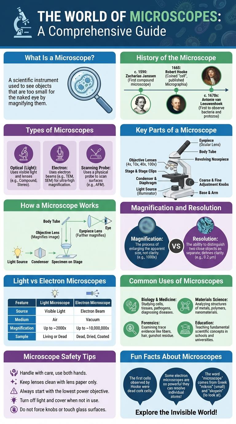
Microscopes magnify tiny objects, revealing intricate details invisible to the naked eye and enhancing scientific discovery. Infographics about microscopes visually simplify complex parts and functions, making information accessible and engaging for learners. Understanding microscope components and uses boosts knowledge in biology, medicine, and materials science.
What Is a Microscope?
| What Is a Microscope? | |
|---|---|
| Definition | A microscope is an optical instrument used to magnify and visualize objects that are too small to be seen clearly with the naked eye. |
| Purpose | To observe the detailed structure of cells, microorganisms, tissues, and other minute specimens for scientific and educational purposes. |
| Types | Common types include optical (light) microscopes and electron microscopes, each suited for different levels of magnification and resolution. |
| Key Components | Includes lenses, light source, stage, eyepiece, and focusing mechanisms that work together to enlarge the image of the specimen. |
| Applications | Used extensively in biology, medicine, material science, and forensic analysis for detailed examination. |
History of the Microscope
The history of the microscope dates back to the late 16th century, with early designs attributed to Dutch spectacle makers Hans Janssen and his son Zacharias. These initial magnifying devices laid the groundwork for modern microscopy.
In 1674, Antonie van Leeuwenhoek pioneered the use of single-lens microscopes, enabling the first observations of microorganisms. Robert Hooke's 1665 publication "Micrographia" introduced detailed drawings of microscopic structures, coining the term "cell." Advancements in lens crafting and illumination techniques throughout the 18th and 19th centuries significantly enhanced microscope performance, fueling scientific discoveries.
Types of Microscopes
Microscopes are essential tools in science that magnify tiny objects invisible to the naked eye. Various types of microscopes serve different purposes depending on the sample and the desired level of detail.
- Light Microscope - Uses visible light to magnify specimens, ideal for viewing cells and tissues.
- Electron Microscope - Employs electron beams for much higher magnification, revealing ultrastructural details of samples.
- Fluorescence Microscope - Utilizes fluorescence to highlight specific components within a specimen, often used in biological and medical research.
Key Parts of a Microscope
The microscope is an essential tool in scientific research, allowing the observation of tiny structures invisible to the naked eye. Understanding its key parts enhances effective use and accurate analysis.
The eyepiece, or ocular lens, magnifies the specimen for viewing. The objective lenses, located on the rotating nosepiece, provide varying levels of magnification for detailed examination.
How a Microscope Works
A microscope magnifies tiny objects using a combination of lenses to enhance details invisible to the naked eye. Light or electrons pass through or bounce off the specimen, creating an enlarged image. This magnified image enables scientists to study cells, bacteria, and other microscopic structures in detail.
Magnification and Resolution
Microscopes are essential tools for viewing objects too small for the naked eye, relying on magnification and resolution to reveal fine details. Magnification enlarges the image, while resolution defines the clarity and ability to distinguish two close points.
- Magnification - The process of enlarging the appearance of an object through lenses or digital zoom.
- Resolution - The microscope's ability to separate two points distinctly, determining image sharpness.
- Impact on Observation - High magnification without adequate resolution results in blurry images, reducing useful detail.
Light vs Electron Microscopes
Microscopes are essential tools in scientific research, allowing the observation of tiny structures invisible to the naked eye. Light microscopes and electron microscopes represent two primary types used for different levels of magnification and detail.
Light microscopes use visible light to illuminate specimens, providing images with color and suitable for live cell observation. Electron microscopes use beams of electrons, offering much higher resolution to reveal ultra-fine structural details at the nanometer scale.
Common Uses of Microscopes
What are the common uses of microscopes? Microscopes are essential tools in scientific research, allowing detailed observation of cells and microorganisms. They are widely used in medical laboratories for disease diagnosis and in educational settings for biology learning.
Microscope Safety Tips
Microscope safety is essential to prevent accidents and preserve the equipment's integrity. Proper handling techniques ensure accurate observations and extend the microscope's lifespan.
- Handle with Care - Always carry the microscope using both hands, supporting the base and arm for stability.
- Keep Lenses Clean - Use lens paper and appropriate cleaning solutions to maintain clear optics.
- Adjust Light Properly - Avoid intense light exposure to protect both the specimen and your eyes.
- Secure Electrical Cords - Prevent tripping hazards by organizing cables away from walkways.
- Store Correctly - Cover the microscope with a dust cover and store it in a dry, safe location after use.
Following these safety tips ensures efficient and safe microscope operation for all users.